3D Modelling of the Coronary Arteries from CT and X-ray Imaging
 |
 |
 |
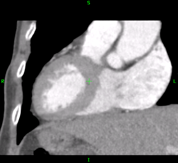
| Coronary CT |
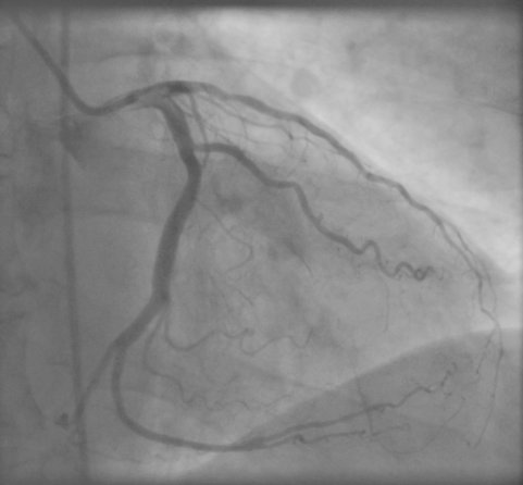
Coronary Angiogram |
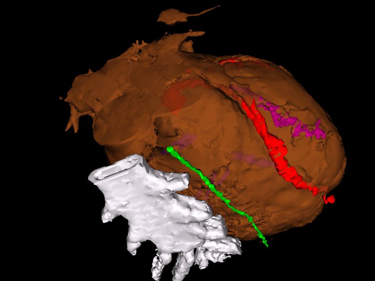
3D Rendering |
Background
A fully four-dimensional model of the heart is useful for analysis
of normal and abnormal cardiac motion. With enough examples it may
be possible to examine the statistics of heart shape variation and
heart motion variation across the population and to characterise
pathology. For image-guided robotic cornary artery bypass there is
also the need to create a fully 4D model of the beating heart that
includes not only the heart muscle (myocardium) but also the
coronary arteries which supply this muscle.
Project Description
In this project we aim to examine two methods that can be used to
produce a 4D model of the coronary arteries. The first is coronary
CT, which gives a fully 3D model at various phases of the cardiac
cycle. This can be segmented to produce a model of the coronary
arteries showing both shape and motion. However, the resolution
and contrast are limited.
Alternatively we can use conventional fluoroscopic X-ray
angiography, which provides high-resolution 2D projections of the
coronary arteries. Two or more such projections can be used to
create a 3D model of the vessel tree if the relative projections
are known and the different arteries can be identified in both
projections.
In this project we will compare the different methods to achieve a
4D model of the beating heart. Methods to achieve 3D
reconstruction from uncalibrated x-rays will be considered. We
will also look at whether registration of CT and X-ray angiograms
would improve the reconstruction. A large database of patients
with coronary CT and multiplanar angiography will be available.
Eddie Edwards
Last modified: Thu Oct 18 16:40:16 BST 2007