exposure of the blot showed that a weak interaction of the A-
and C-subunits with the EF2 and EF1/EF2 double mutants
could still be detected (Fig. 3C, OE). The combination of intact
cell data and in vitro binding assays provide evidence that PR70
is a calcium binding protein and that interaction with the core
dimer of PP2A is enhanced by binding of calcium to the second
EF-hand motif. Calcium does not affect interaction of PR70
with Cdc6 but increases the association of the PP2A core dimer
with Cdc6 in a manner dependent upon binding of calcium to
the second EF-hand of PR70.
units identified a conserved domain in the central region of
PR70 (supplemental Figs. S1
and PR72 (PPP2R3A). A series of truncation mutants were con-
structed to identify regions within PR70 that were important
for interaction with PP2A and Cdc6. FLAG-tagged mutants
were expressed in COS-7 cells and immunoprecipitated with
anti-FLAG antibody. The ability of the mutants to incorporate
into endogenous PP2A heterotrimers was determined by
immunoblotting for associated A- and C-subunits. The
unique region and interacted with endogenous PP2A subunits
to the same extent as full-length PR70 (Fig. 4B). Deletion of a
(
domain,
amino acids 125 and 136 of PR70 were necessary for interaction
with the PP2A core dimer.
proteins from humans to flies (Fig. 5A). The role of the FYF
motif was tested by mutating these residues to alanines (Fig. 5B)
and determining the effects on interaction with the AC core
dimer. Mutation of any one of these residues resulted in a sig-
nificant loss of interaction with endogenous PP2A (Fig. 5C). A
longer exposure of the immunoblot showed that small amounts
of the A- and C-subunits could be detected in immunoprecipi-
tates of each of the mutants (not shown). Although the interac-
tion of the FYF mutants was severely compromised in intact
cells, these mutants still bound to PP2A when assayed in vitro
by GST pulldown experiments (not shown). These results indi-
cate that the FYF motif contributes to the interaction of PR70
with the A-subunit.
ments with GST-Cdc6 were performed with full-length PR70,
the
)
pulldown
pulldown
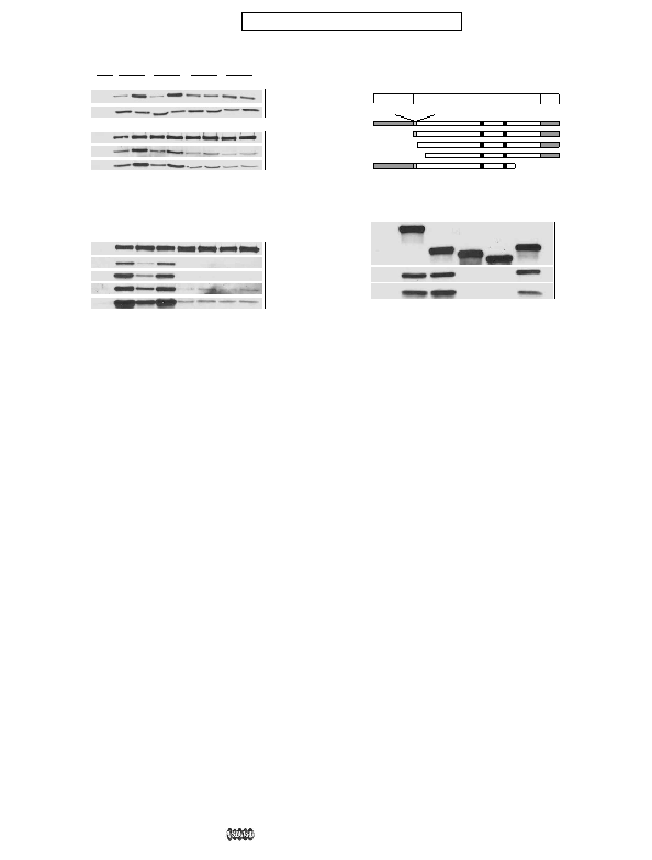
IP
EF2. FLAG-PR70 and the indicated EF-hand mutants were transiently
expressed in COS-7 cells, and the cells were lysed in standard lysis buffer (N) or
lysis buffer containing EGTA (E) or CaCl
teins were detected by immunoblotting with anti-FLAG (FL-PR70), anti-A-sub-
unit (A), and anti-C-subunit (C) antibodies. B, GST pulldown assays were per-
formed using immobilized GST-Cdc6 as described for A. Lane 1 of panels A and
B shows a control pulldown assay using GST alone. C, COS-7 cells were tran-
siently transfected with FLAG-tagged wild-type PR70 or the indicated EF-
hand mutants, and lysates prepared with standard buffer were immunopre-
cipitated with anti-FLAG antibody. The immunoprecipitates were resolved by
SDS-PAGE and immunoblotted as described in A. Overexposures of the anti-
A-subunit (A OE) and anti-C-subunit (C OE) immunoblots are also shown.
C
N3
N2
N1
125-575
136-575
162-575
1-441
IP
gram of PR70 showing the region containing the conserved R3 domain and
PR70-unique regions. The truncation mutants used in binding assays are
shown below with their corresponding designations on the left and amino
acid numbers on the right. The conserved FYF (FYF motif) and EF-hand motifs
(EF1 and EF2) are also depicted. B, interaction of PR70 truncation mutants with
the A- and C-subunits of PP2A. FLAG-tagged PR70 (PR70) and the indicated
truncation mutants were transiently expressed in COS-7 cells. The cells were
lysed and the FLAG-tagged proteins immunoprecipitated with anti-FLAG
antibody (Anti-FLAG IP). The immunoprecipitates were resolved by SDS-PAGE
and immunoblotted with anti-FLAG (FLAG), anti-A-subunit (A), and anti-C-
subunit (C) antibodies. A control immunoprecipitate using lysate from cells
transfected with the empty expression vector (Emp Vec) is shown in the first
lane.