Cdc6). Similar results were seen in U2OS cells. These data indi-
cate that knockdown of PR70 results in an increase in the levels
of endogenous and exogenous Cdc6 that is dependent on the
presence of phosphorylatable residues at the N-terminal phos-
phorylation sites.
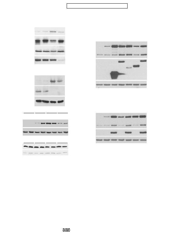
plasmids containing the CMV promoter, FLAG-tagged PR70
was expressed at levels 5- to 10-fold higher than the endoge-
nous protein (not shown). Co-expression of CDK2 and Cdc6
caused a substantial increase in Cdc6 levels as reported previ-
ously (15). Expression of wild-type PR70 caused an increase in
the levels of both co-transfected and endogenous Cdc6 (Fig.
8A). Expression of the
increased the levels of Cdc6. In contrast, expression of the
mock transfected (Mock) or transfected with control (Luc), PP2A catalytic subunit
(PP2A-C), or PP5 (PP5) siRNA. Cells were harvested 48 h after transfection and
immunoblotted with anti-Cdc6, PP2A-C, PR70, or PP5 antibodies. B, HeLa cells
were mock transfected (Mock) or transfected with control (Luc), PR70-1, or PR70-2
siRNA. Cells were harvested 48 h after transfection and immunoblotted with anti-
Cdc6, PR70, or actin (as a loading control) antibodies. C, HeLa cells were co-trans-
fected with control or PR70-1 siRNA and plasmids encoding GFP-tagged versions
of wild type Cdc6 (wtCdc6) or Cdc6 in which the N-terminal phosphorylation sites
were mutated to aspartic acid (DDD-Cdc6) or alanine (AAA-Cdc6). Forty-eight
hours later, the cells were harvested and lysates were analyzed by immunoblot-
ting with antibodies against Cdc6 or glyceraldehyde-3 phosphate dehydrogen-
ase (GPDH) as a loading control. D, duplicate samples of the lysates described in
B, from cells co-transfected with Cdc6 and either luciferase control (Luc siRNA) or
PR70-1 (PR70) siRNAs were immunoblotted with anti-PR70 antibodies to confirm
knockdown of PR70.
cells were transfected with empty vector, or plasmids encoding GFP-Cdc6
(Cdc6), myc-tagged CDK2 (CDK2), or FLAG-tagged constructs of wild-type
PR70 (PR70) or the indicated PR70 mutants. Cells were harvested 24 h after
transfection, and lysates were analyzed by immunoblotting with antibodies
against Cdc6, actin, FLAG, or myc as indicated at the right. The Cdc6 antibod-
ies detected both the expressed Cdc6 (GFP-Cdc6) and endogenous Cdc6
(Endo-Cdc6). B, U2OS cells were co-transfected with empty vector or plasmids
expressing FLAG-PR70 and plasmids expressing GFP-tagged versions of wild-
type Cdc6 (wtCdc6), or the AAA (AAA-Cdc6), or DDD (DDD-Cdc6) triple phos-
phorylation site mutants of Cdc6. Cells were harvested 24 h later, and lysates
were analyzed by immunoblotting with antibodies against Cdc6, actin, or the
FLAG epitope as indicated at the right.