Atrial wall thickness
Pulmonary vein isolation (PVI) is often the first procedure performed in patients referred for catheter ablation of atrial fibrillation (AF). The procedural success rate of an index PVI varies between 15-75%. An important cause for this could be non-transmural lesion formation due to insufficient catheter tip-tissue contact.
Recent developments have enabled measurement of the force with which the catheter tip touches the myocardium. However, in order to determine the optimal RF power and application time, local myocardial thickness also needs to be known. Non-invasive 3-dimensional imaging using cardiac computed tomography (CCT) can accurately provide information about the left atrium (LA) wall thickness. New sequence design has also enabled acquisition of the LA wall thickness using cardiac magnetic resonance (CMR). The reliability of these parameters need to be determined prior to evaluating the clinical utility.
MRI (n=10)
The MRI datasets can be downloaded directly along with their manual labellings
All images were acquired on a Philips 3T Achieva, with 3D FLASH, typical FOV: 280 x 190 x 120 mm, 1.4-mm isotropic resolution, FA: 20°, TE/TR/TI: 2.7/5.9/120 ms. Respiratory gating was achieved using pencil-beam navigator and the average scan time was 12 min. Cardiac triggering was used to ensure data acquisition was carried out in mid atrial diastole
Please refer to this SCMR abstract for further details
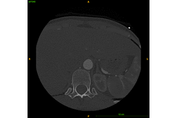
CT (n=10)
The CT datasets can be downloaded along with their manual labellings on this dataset of the atria wall and chamber.
Coronary CTA was performed on a Philips 256 iCT scanner. All paients were injected with an intravenous contrast agent. The scans were ECG-gated in a single breath hold. The images were reconstructed to a 0.8 to 1 mm slice thickness, with a 0.4 mm slice increment and a 250 mm field of view. The image matrix was kept at 512 × 512 matrix, constructed with and a sharp reconstruction kernel.