Interaction of the lipid acyl chains with AQP0
In contrast to the headgroups, acyl chains seem to be the
crucial element guiding the interaction of annular lipids with
the protein. Comparison of the lipids in AQP0EPL and
AQP0DMPC reveals that the acyl chains in the extracellular
leaflet occupy quite similar positions. The acyl chains of PE3
and PE4 extend along the same paths as those of the
corresponding lipids, PC3, and PC4. The two acyl chains of
PE2 are in similar positions as those occupied by one of the
acyl chains of PC1 and one of the acyl chains of PC2
The positions of the acyl chains in the cytoplasmic leaflet
vary to a greater degree between AQP0EPL and AQP0DMPC
than those of the acyl chains in the extracellular leaflet
is narrower on the extracellular side of the bilayer, which may
force the lipids in this leaflet into more defined positions than
those in the cytoplasmic leaflet. This idea is supported by the
average B-factors of the lipids, which tend to be lower for the
lipids in the extracellular leaflet, suggesting that their mobi-
lity is more restricted compared with those in the cytoplasmic
Lipid PE7 in AQP0EPL illustrates how the protein surface
defines the positions of the acyl chains of annular lipids
surface of AQP0 that guides it straight towards the centre of
the bilayer. The other acyl chain runs in between the bulky
side chains of Trp 10 and Phe 18 and then bends sharply to
evade the side chain of Leu 95, thus filling in a cleft formed by
Trp 10 on one side and Val 91 and Leu 95 on the other. The
two acyl chains of PE6 extend over the surface of this cleft,
completely covering it. Phosphatidylcholine 7, the lipid in
AQP0DMPC that corresponds to PE7 in AQP0EPL, adapts
with PE7, the glycerol backbone of PC7 is shifted by about
3A
° away from the centre of the bilayer, which is probably
caused by the bulky side chain of Phe 14 adopting a different
conformation in AQP0DMPC. One of the acyl chains of PC7
follows the same groove in the AQP0 surface as the corre-
sponding acyl chain of PE7, extending straight to the centre of
the bilayer. The second acyl chain, however, follows a gap in
between the side chains of Trp 10 and Phe 14 in its different
position. It then immediately fills the cleft formed by residues
Trp 10, Phe 14, Val 91, and Leu 95. Thus, both PE7 and
PC7 adopt conformations that allow the acyl chains to fill in
the large hydrophobic pocket in the AQP0 surface, but in
different ways. To even out the protein surface is a crucial
function of annular lipids, because it ensures that the bilayer
forms a tight seal around the membrane protein and
preserves the separation of the cellular interior from the
external environment.
Discussion
Comparison of our new AQP0EPL structure with the pre-
viously determined AQP0DMPC structure shows that the an-
nular lipids studied here have very little influence on the
structure of AQP0. With only a few exceptions, such as Phe
14, the amino-acid residues in the AQP0EPL and AQP0DMPC
structures, in particular the ones lining the water channel,
have nearly identical conformations. The virtually identical
channel structure in AQP0EPL and AQP0DMPC is consistent
with a previous study that showed that the lipid environment
although only a very limited range of lipid compositions were
tested in this study.
Electron crystallography of AQP0 2D crystals makes it
possible to visualize the lipids surrounding the membrane
protein. In our studies, only a single layer of lipid molecules
separates the individual tetramers in the 2D crystals
(Figure 2A). This tight packing of the lipids in the 2D crystals
restricts their mobility and makes it possible to see the lipids
in the density maps, providing us with the opportunity to
study the interactions of annular lipids with AQP0. However,
this situation is not typical for a biological membrane, in
which membrane proteins tend to be separated by a larger
number of lipids. Nevertheless, the 2D crystals formed
in vitro display the same lattice parameters as the 2D arrays
tion of the AQP0 arrays in the lens membrane. Furthermore,
AQP0 forms 2D crystals with lipids as different as DMPC and
EPLs. The sandwiching between two adjacent tetramers may
thus limit the mobility of the lipids but may not impose
further restrictions on the behaviour of the lipids surrounding
AQP0. Our findings concerning the interaction of annular
lipids with AQP0 in the 2D crystals should therefore bear
relevance for membrane proteins surrounded by a larger
number of lipids. Nevertheless, further studies will be
required to confirm this notion.
The structures of AQP0 in two different lipid environments
allow us to see how the protein and the lipid bilayer adapt to
each other. To accommodate proteins in a lipid bilayer with a
different hydrophobic thickness, a situation known as hydro-
phobic mismatch, it has been proposed that either the lipids
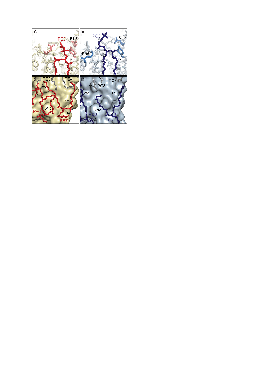
Figure 4 EPL headgroups and acyl chains. (A) Lipid PE3 interacts
with AQP0 through a putative lipid-binding motif formed by Arg196
and Tyr105. (B) In AQP0DMPC the same residues do not interact with
the corresponding lipid, PC3. (C) Lipid PE7 in AQP0EPL bends to
follow the surface features of AQP0 and fills in a pocket formed by
residues Trp10, Val91, and Leu95. (D) The corresponding lipid in
AQP0DMPC, PC7, follows a different path but fills in the same pocket
in the AQP0 surface.
Interaction of AQPO with E. coli lipids
RK Hite et al
The EMBO Journal
VOL 29 | NO 10 | 2010
&
2010 European Molecular Biology Organization
1656